Tendon Diagram / They are remarkably strong, having one of the highest tensile strengths found among soft tissues.. Superficial posterior muscles of the forearm posterior compartment muscles of the forearm. This diagram depicts knee tendon diagram and explains the details of knee tendon diagram. Tendons are found throughout the body, from the head and neck all the way down to the feet. Human anatomy for women 12 photos of the human anatomy for women human anatomy for bds 1st year, human anatomy for drawing, human anatomy for dummies, human anatomy for nurses ppt, human anatomy for sketching pdf, human muscles, human anatomy for bds 1st year, human anatomy for drawing, human anatomy for. On the other hand, the insertion is where a tendon attaches that muscle to the *more* movable bone.
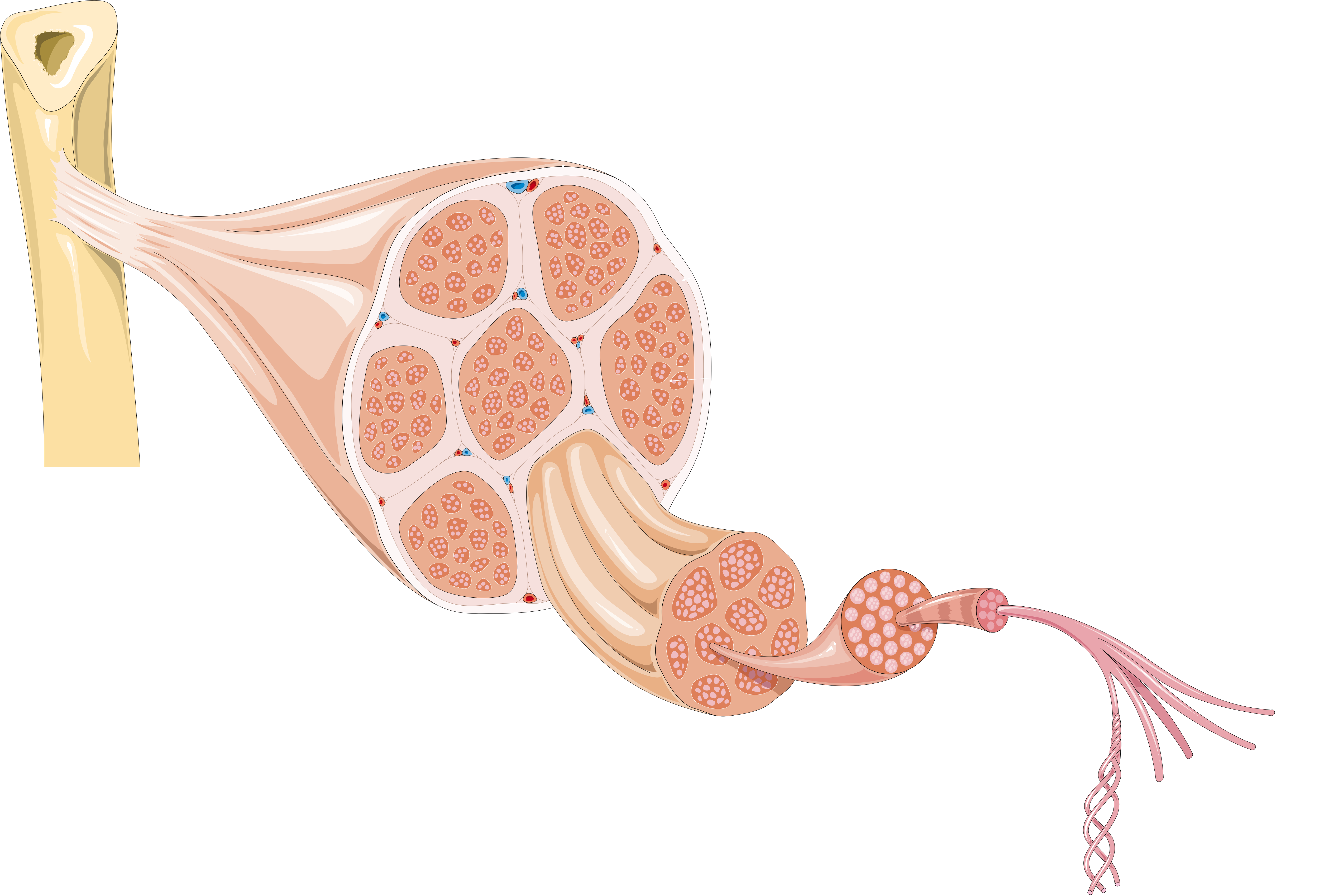
Superficial posterior muscles of the forearm posterior compartment muscles of the forearm. Related posts of muscles and tendons of the leg muscle anatomy gluteus. The fascicle contains the basic fibril of the ligament or tendon, and the fibroblasts, which are the biological cells that produce the ligament or tendon. A muscle's origin is where a tendon attaches it to the *less* movable bone. The achilles tendon is the largest.
Ligaments Tendons And Muscles Of The Hip Joint Naples Best Hip Surgeon from zehrcenter.b-cdn.net Fall on one point of shoulder and can rupture these ligaments with dislocation of ac joint. The ecu tendon works along with the ecrl and ecrb to straighten the wrist. Biceps and triceps tendon rupture. Diagram depicting the bones, ligaments and muscles throughout the hand and fingers. The coracobrachialis muscle lies deep to the biceps brachii in the arm. Attaches the calf muscles to the calcaneus, most important muscles for running, jumping, walking etc. The ligament or tendon then is split into smaller entities called fascicles. Tendon, tissue that attaches a muscle to other body parts, usually bones.
Allows the action of raising the foot.
2 ligaments (trapezoid& conoid ligaments) attach the clavicle coracoid process of scapula these tiny ligaments (w/ acominoclavicular joint) keep scapula attached to clavicle. Bones, cartilage, ligaments, and tendons. Top (dorsal) view of foot & ankle number 1 and 2: Here you can see the tendons that extend down the top of your. If you tear the biceps tendon at the shoulder, you may lose some strength in your arm and have pain when you forcefully turn your arm from palm down to palm up. Diagram of the shoulder, including the location of the rotator cuff. Arguably, the most important tendon is the achilles tendon, which allows the calf muscles to move the ankle joint. Limit plantar flexion resist adduction limit dorsi flexion. Your biceps tendons attach the biceps muscle to bones in the shoulder and in the elbow. The achilles tendon attaches the muscles of the calves to the bones of the ankle and foot. Muscle anatomy gluteus 12 photos of the muscle anatomy gluteus gluteus muscle anatomy ct, gluteus muscle anatomy mri, human muscle anatomy gluteus maximus, muscle anatomy gluteus, muscle anatomy of gluteal, human muscles, gluteus muscle anatomy ct, gluteus muscle anatomy mri, human muscle anatomy gluteus maximus. Diagram depicting the bones, ligaments and muscles throughout the hand and fingers. Learn about these muscles, their origin and insertion points, and their functional anatomy.
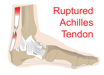
The achilles tendon attaches the muscles of the calves to the bones of the ankle and foot. The calf muscles gastrocnemius and soleus which are connected to the calcaneus via the achilles tendon. The fascicle contains the basic fibril of the ligament or tendon, and the fibroblasts, which are the biological cells that produce the ligament or tendon. The rotator cuff is a group of four muscles and tendons that surround the glenohumeral joint. The largest of these shoulder muscles is the.
Achilles Tendon Ruptures from www.washingtonheelpain.com Tendons transmit the mechanical force of muscle contraction to the bones. If you tear the biceps tendon at the shoulder, you may lose some strength in your arm and have pain when you forcefully turn your arm from palm down to palm up. Fall on one point of shoulder and can rupture these ligaments with dislocation of ac joint. Ligaments connect bones to each other to support a joint. These structures work together to support the body, enable a range of movements, and send messages from the brain to. A tendon is a band of tissue that connects a muscle to a bone. The rotator cuff is a group of four muscles and tendons that surround the glenohumeral joint. Tendons attach muscles to bones.
Ligaments join the knee bones and provide stability to the knee:
Related posts of muscles of the lower back and hip diagram human anatomy for women. Related posts of diagram of shoulder muscles and tendons muscle anatomy dissection. Biceps and triceps tendon rupture. The changes in ligaments and tendons generally occur more slowly than adaptation in bone, because ligaments and tendons have less vascular supply. The anterior cruciate ligament prevents the femur from sliding backward on the tibia (or the tibia sliding forward on the femur). Ligaments and tendons are adapted in response to changes in mechanical stiffness. Human anatomy for women 12 photos of the human anatomy for women human anatomy for bds 1st year, human anatomy for drawing, human anatomy for dummies, human anatomy for nurses ppt, human anatomy for sketching pdf, human muscles, human anatomy for bds 1st year, human anatomy for drawing, human anatomy for. Ligaments connect bones to each other to support a joint. The largest structure in the above schematic is the tendon (shown) or the ligament itselt. 2 ligaments (trapezoid& conoid ligaments) attach the clavicle coracoid process of scapula these tiny ligaments (w/ acominoclavicular joint) keep scapula attached to clavicle. It attaches to the wrist bone, the pisiform, and as well as the 5th hand bone. Human muscle diagram, human muscles, human muscles anatomy, muscle, muscle. A foot pain diagram is a great tool to help you work out what is causing your ankle and foot pain.
Groin strain treatment rehabilitation exercises although there is often swelling oedema as a result of a groin strain this is often not visible to the eye groin strains are graded 1 2 or 3 depending on the extent of the injury groin muscle diagram diagram muscles in groin area male groin muscle diagram diagram muscles in groin area male anatomy groin human photo groin. Tendons attach muscles to bones. It attaches to the wrist bone, the pisiform, and as well as the 5th hand bone. The tendon travels along the inside of the forearm on the side of the small finger and crosses the wrist. It is constructed in such a way that we can move the arms to.
File Tendon Anatomy 1 Smart Servier Png Wikimedia Commons from upload.wikimedia.org The shoulder joint is formed the rotator cuff is a collection of muscles and tendons that. Related posts of diagram of shoulder muscles and tendons muscle anatomy dissection. The knee joint is a complex structure that involves bones. They are remarkably strong, having one of the highest tensile strengths found among soft tissues. Numerous muscles help stabilize the three joints of. A foot pain diagram is a great tool to help you work out what is causing your ankle and foot pain. The rotator cuff is a group of four muscles and tendons that surround the glenohumeral joint. Tendons are found throughout the body, from the head and neck all the way down to the feet.
The achilles tendon attaches the muscles of the calves to the bones of the ankle and foot.
Again, our knowledge of how mechanical stimulus mediates ligament and tendon structure is more empirical and less. A tendon is a band of tissue that connects a muscle to a bone. Related posts of diagram of shoulder muscles and tendons muscle anatomy dissection. This tendon connects the patella (kneecap) to the tibia. The fascicle contains the basic fibril of the ligament or tendon, and the fibroblasts, which are the biological cells that produce the ligament or tendon. Flexor tendon lacerations are classified into five zones 2, 15, 16. The coracobrachialis muscle lies deep to the biceps brachii in the arm. The two peroneal tendons in the foot run side by side behind the outer ankle bone. A muscle's origin is where a tendon attaches it to the *less* movable bone. Hand a hand is a prehensile multi fingered appendage located at the end of the forearm or forelimb of primates such as humans chimpanzees monkeys and lemurs human anatomy for the artist the dorsal hand the dorsal the easiest tendons to identify in the dorsal hand are those of the extensor digitorum muscle its name means extensor of the digits which is Diagram showing the tendons and ligaments of the ankle and. Bones, cartilage, ligaments, and tendons. A foot pain diagram is a great tool to help you work out what is causing your ankle and foot pain.


Posting Komentar
0 Komentar